
When someone experiences foot or ankle pain, it’s not always clear what the cause might be. Sometimes it’s a recent injury, like a sprain or possible fracture. Other times it may be pain that’s been building for months without a clear explanation. In both situations, getting an accurate diagnosis is the first and most important step toward finding the right treatment. This is where high-resolution digital X-rays come into play—especially in modern clinics like Gelbmann Podiatry, where advanced imaging technology is part of the standard of care.
In the past, traditional X-rays provided a basic view of bones and joints. While useful, they often lacked the detail needed to identify more subtle problems. Today, digital X-rays have transformed the diagnostic process, giving podiatrists a much clearer and faster look inside the body. For foot and ankle care, this means quicker answers, more accurate diagnoses, and better outcomes for patients.
Table of Contents
Why Imaging Matters in Foot and Ankle Care
The human foot is made up of 26 bones, 33 joints, and more than 100 muscles, tendons, and ligaments. It’s a small but complex structure, and when something goes wrong, even a minor misalignment or injury can cause significant pain. Because so many issues in the feet and ankles occur beneath the surface—inside joints, around tendons, or along the bones—visual examination alone isn’t enough to diagnose them accurately.
Podiatrists rely on imaging tools to “see inside” the foot and understand what’s really happening. Whether a patient is dealing with a suspected fracture, a bunion, a bone spur, or an unknown source of pain, X-rays help paint a clearer picture. The quality of that picture can make a major difference in how quickly and effectively the problem is treated.
What Makes Digital X-Rays Different?
Digital X-rays use advanced sensors instead of traditional film to capture images of the body. These sensors immediately transmit high-resolution images to a computer screen, allowing podiatrists to view, enlarge, and analyze them in real time. The images are crisper, more detailed, and easier to manipulate, which allows for a more precise look at the structure of the foot and ankle.
Because the process is digital, there is no waiting for film to develop. This means patients can get immediate feedback and diagnosis during their visit, often within minutes. The clarity of digital imaging also helps detect small changes in bone structure, subtle fractures, or early signs of joint degeneration—things that might be missed with older imaging systems.
At Gelbmann Podiatry, high-resolution digital X-rays are a core part of the diagnostic process. By having this technology on-site, the clinic can deliver faster care and better results, reducing the need for outside imaging appointments and unnecessary delays in treatment.
Diagnosing Common Foot and Ankle Conditions
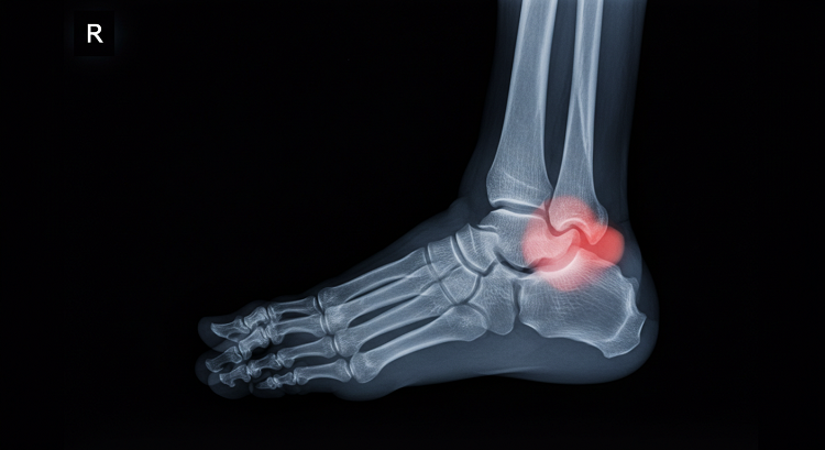
One of the most common uses of digital X-rays in podiatry is to evaluate for fractures. Foot and ankle injuries can occur from sports, falls, or even something as simple as stepping the wrong way off a curb. In these cases, high-resolution X-rays allow doctors to identify small bone breaks that may not be obvious through a physical exam alone.
Another frequent use is in the diagnosis and monitoring of bunions, which involve a bony bump at the base of the big toe joint. X-rays can help determine the severity of the deformity, show how the bones are aligned, and guide decisions about treatment—whether that’s conservative care or surgical correction.
Digital X-rays are also valuable for diagnosing conditions like arthritis, where joint space narrowing and bone changes may indicate inflammation or degeneration. With clearer images, podiatrists can better assess the progression of the disease and tailor treatment to the patient’s needs.
Bone spurs, heel pain, alignment problems, and other structural abnormalities are also commonly diagnosed through X-ray. In some cases, the issue may be related to previous injuries that have healed improperly or anatomical problems that worsen over time. Imaging plays a key role in understanding these deeper issues.
Benefits for the Patient
One of the most noticeable benefits of digital X-rays is the speed of the process. Instead of waiting for results from a separate imaging center, patients can receive their diagnosis and begin treatment all in one visit. This reduces anxiety, shortens recovery time, and helps people get back to their daily activities faster.
Digital X-rays also use significantly less radiation than traditional film X-rays, making the procedure safer for patients. Since only the specific area being examined is exposed, the rest of the body remains protected. This is especially important for patients who require repeated imaging over time, such as those with chronic conditions.
Another benefit is the ability to store and retrieve images electronically. Digital records make it easier for podiatrists to track changes over time, compare current and past images, and share information with other healthcare providers if needed. This leads to better communication, more coordinated care, and improved long-term outcomes.
Supporting Personalized Treatment Plans
Every foot is different, and so is every patient. With high-resolution digital X-rays, podiatrists can create treatment plans that are based on a detailed, accurate view of the patient’s unique anatomy. Whether the goal is to relieve pain, improve mobility, or correct a deformity, having the right imaging tools supports smarter, more personalized decisions.
For example, a patient with heel pain may assume it’s due to plantar fasciitis, a common condition. But a digital X-ray might reveal a bone spur or a stress fracture that needs a different approach. Without imaging, these conditions could be missed—or mistreated—leading to frustration and delays in healing.
At clinics like Gelbmann Podiatry, where patient-focused care is a priority, access to digital imaging allows the team to move quickly from diagnosis to treatment, ensuring that each patient receives the care that’s best suited to their situation.
The field of podiatry continues to evolve, with better tools and techniques making foot and ankle care more precise and effective than ever before. Digital X-rays are a prime example of this progress. By delivering clearer images with less radiation and faster results, they help both doctors and patients make smarter decisions and see better outcomes.
Whether someone is dealing with a recent injury, chronic pain, or an ongoing issue that hasn’t responded to treatment, digital imaging is often the key to uncovering the cause and moving forward with confidence.
Clinics like Gelbmann Podiatry are leading the way by combining state-of-the-art technology with personalized care. For patients, this means access to faster, safer, and more effective foot and ankle diagnostics—all under one roof. In a field where every step counts, digital X-rays offer a clearer path to relief and recovery.